Showing posts with label brain damage. Show all posts

Monday, December 10, 2012

Brain pacemaker delivers constant Deep-Brain Stimulation (DBS) for Alzheimer’s patients


Credit Image: Functional Neuromodulation

Once implanted into the brain, a pacemaker-like device delivering electrical stimulation could help improve the memory of Alzheimer’s patients. Technology Review reports.

Using electrodes, Deep-Brain Stimulation (DBS) is already used to treat patients with Parkinson’s, epilepsy, and obsessive-compulsive disorder.

In this Alzheimer’s trial, co-chaired by Constantine Lyketsos of Johns Hopkins, the device was placed into a region of the brain involved in learning and memory.

In Alzheimer’s patients, brain tissue atrophies and the reduction of memory and thinking skills increase over time.

According to the Johns Hopkins team, electrical shocks could stimulate critical neural networks disrupted by Alzheimer’s.

Recent trials for potential Alzheimer’s drugs have failed to halt or stave off cognitive decline.

Now, in a pilot study with these deep brain stimulators, after one year of constant stimulation, brain scans of six Alzheimer’s patients showed signs of increased neuron activity in areas involving learning and memory, although, it could be unlikely to actually reverse the effects or damage to the brain caused by Alzheimer’s disease.

The researchers are recruiting patients into the new trial initiated by Toronto-based Functional Neuromodulation. The trial will track patients who have the device for a year using doctor observations and brain scans.

[Via Technology Review]


Monday, August 13, 2012

Leukoaraiosis: 'Harmless' condition shown to alter brain function in elderly

Researchers at the Mayo Clinic say a common condition called leukoaraiosis, made up of tiny areas in the brain that have been deprived of oxygen.

They appear as bright white dots on MRI scans, is not a harmless part of the aging process, but rather a disease that alters brain function in the elderly.

Results of their study are published online in the journal Radiology. "There has been a lot of controversy over these commonly identified abnormalities on MRI scans and their clinical impact," said Kirk M. Welker, M.D., assistant professor of radiology in the College of Medicine at Mayo Clinic in Rochester, Minn.

"In the past, leukoaraiosis has been considered a benign part of the aging process, like gray hair and wrinkles."

 Leukoaraiosis, also called small vessel ischemia and often referred to as unidentified bright objects or "UBOs" on brain scans, is a condition in which diseased blood vessels lead to small areas of damage in the white matter of the brain.

The lesions are common in the brains of people over the age of 60, although the amount of disease varies among individuals.

"We know that aging is a risk factor for leukoaraiosis, and we suspect that high blood pressure may also play a role," Dr. Welker said.

Dr. Welker's team performed functional MRI (fMRI) scans on cognitively normal elderly participants recruited from the Mayo Clinic Study of Aging between 2006 and 2010.

In 18 participants, the amount of leukoaraiosis was a moderate 25 milliliters, and in 18 age-matched control participants, the amount of disease was less than five milliliters.

The patients were imaged in an MRI scanner as they performed a semantic decision task by identifying word pairs and a visual perception task that involved differentiating straight from diagonal lines.

fMRI is a special type of magnetic resonance imaging that measures metabolic changes in an active part of the brain.

Although both groups performed the tasks with similar success, the fMRI scans revealed different brain activation patterns between the two groups.

Compared to members of the control group, patients with moderate levels of leukoaraiosis had atypical activation patterns, including decreased activation in areas of the brain involved in language processing during the semantic decision task and increased activation in the visual-spatial areas of the brain during the visual perception task.

"Different systems of the brain respond differently to disease," Dr. Welker explained. "White matter damage affects connections within the brain's language network, which leads to an overall reduction in network activity."

He pointed out that identifying leukoaraiosis in the brain is important, both for individual patients undergoing brain mapping for surgery or other treatments and for research studies.

 For improved neurological health, Dr. Welker said efforts should be taken to prevent leukoaraiosis from occurring.

"Our results add to a growing body of evidence that this is a disease we need to pay attention to," he said.

"Leukoaraiosis is not a benign manifestation of aging but an important pathologic condition that alters brain function."

Friday, June 15, 2012

Anti-anxiety Drug calms fears by altering brain chemistry

An advance in understanding the brain’s fear circuitry has been revealed by a research team. They say it may hold particular promise for people at risk for anxiety disorders, including those suffering post-traumatic stress disorder (PTSD). Findings are reported in the journal Molecular Psychiatry.

“What is most compelling is our ability to translate first from mice to human neurobiology and then all the way out to human behaviour,” says Ahmad Hariri, a neurobiologist at Duke University. “That kind of translation is going to define the future of psychiatry and neuroscience.”

The common thread in their studies is a gene encoding an enzyme called fatty acid amide hydrolase, or FAAH.

The enzyme breaks down a natural endo-cannabinoid chemical in the brain that acts in essentially the same way that Cannabis, aka marijuana, does (hence the name endo-cannabinoid).

Earlier studies had suggested that blocking the FAAH enzyme could decrease fear and anxiety by increasing endo-cannabinoids, which is consistent with the decreased anxiety some experience after smoking marijuana.

In 2009, Hariri’s lab found that a common variant in the human FAAH gene leads to decreased enzyme function with affects on the brain’s circuitry for processing fear and anxiety.

In the new study, Andrew Holmes’ group at the National Institute on Alcoholism and Alcohol Abuse tested the effects of a drug that blocks FAAH activity in fear-prone mice that had also been trained to be fearful through experiences in which they were delivered foot shocks.

Tests for the ability of those mice to get over their bad experiences found that the drug allowed a faster recovery from fear thanks to higher brain endo-cannabinoid levels.

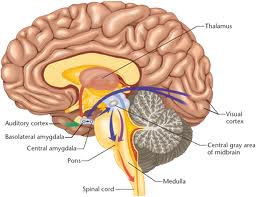
More specifically, the researchers showed that those drug effects traced to the amygdala, a small area of the brain that serves as a critical hub for fear processing and learning.

To test for the human relevance of the findings, Hariri’s group went back to the genetic variant they had studied earlier in a group of middle-aged adults.

They showed study participants a series of pictures depicting threatening faces while they monitored the activity of their amygdalas using functional magnetic resonance imaging (fMRI) scans. They then looked for how the genetic variant affected this activity.

While the activity of the amygdala in all participants decreased over repeated exposures to the pictures. But people who carried the version of the FAAH gene associated with lower enzyme function and higher endo-cannabinoid levels showed a greater decrease in activity.

Hariri says that suggests that those individuals may be better able to control and regulate their fear response.

Further confirmation came from an analysis led by Duke’s Avshalom Caspi and Terrie Moffitt of 1,000 individuals in the Dunedin Study, who have been under careful observation since their birth in the 1970s in New Zealand.

Consistent with the mouse and brain imaging studies, those New Zealanders carrying the lower-expressing version of the FAAH gene were found to be more likely to keep their cool under stress.

“This study in mice reveals how a drug that boosts one of the brain’s naturally occurring endo-cannaboids enables fear extinction, a process that forms the basis of exposure therapy for PTSD,” Holmes says.

“It also shows how human gene variation in the same chemical pathways modulates the amygdala’s processing of threats and predicts how well people cope with stress.”

Studies are now needed to further explore both the connections between FAAH variation and PTSD risk as well as the potential of FAAH inhibition as a novel therapy for fear-related disorders, the researchers say.

More news from Duke University: http://today.duke.edu/

Tuesday, April 3, 2012

Building brains: toward a do-it-yourself guide

With an estimated 100 billion neurons chattering to one another through perhaps a quadrillion synaptic connections, the human brain has the most intricately complicated structure and function of any object in the known universe.

How do you build something like that? In fact, how does something so ferociously complex start off as a single cell and then create itself?

The problem might at first seem impossible to fathom, even with the knowledge that genetics, natural selection, and a half billion years of evolution can accomplish amazing things. But complexity does not have to be born of complexity, as mathematics and computer programs routinely demonstrate.

Very simple equations can give rise to the stunning complexity of fractals, for example.

Several recent neuroscience studies may be illuminating some of the structural and development principles that organize the brain. Those principles include:
  • Co-opting older, simpler genetic programs and elaborating on them to new effect.
  • Maintaining geometric order.
  • Relying on hierarchies of control.
True, science is still very far from knowing enough to define confidently how a brain takes shape (let alone how one works). Yet it’s still fascinating to see how much structure may be determined by relatively simple rules of thumb.

Rule 1: Elaborate on older genetic mechanisms
In the earliest stages of embryonic development, the nervous systems of vertebrate animals are much like those of simpler invertebrates such as worms and starfish.

The big differences start to appear as a result of actions by certain parts of the vertebrate embryo’s body called signaling centers.

These centers release proteins that tell portions of the embryo’s exterior layer of tissue (the neuroectoderm) to organize themselves into major divisions of what will eventually become brain and spinal cord.

The standing inference has been that the genes for those signaling proteins emerged as part of whatever evolutionary changes first split the vertebrate and invertebrate lines.

New work by Ariel M. Pani of the University of Chicago and others, recently published in Nature, suggests that is not the case, however.

The acorn worm Saccoglossus kowaleskii. (Credit: Ariel Pani)

Pani and her colleagues have identified a highly similar set of genes active in the acorn worm (Saccoglossus kowalevskii), a tiny aquatic invertebrate.

According to their studies, not only do these genes have sequences that echo those of the vertebrates but they also express themselves in a similar pattern and in corresponding parts of the body.

They also help to organize the formation of features in the worms’ ectodermal tissue layer.

What’s most curious about Pani’s findings — and makes them controversial — is that the acorn worms fall into the hemichordate branch of the invertebrate family, and as such are much more distant cousins to vertebrates than are many other creatures (such as sea squirts) that seem to lack these signaling center genes.

The implication is that the genes were present in some common wormy ancestor from about 500 million years ago but that the other lines of related invertebrates subsequently lost them during evolution.

Yet some biologists question both whether the genes are truly missing from the other invertebrates and whether they really serve to organize the acorn worms’ ectoderm to the sophisticated degree claimed. (Katherine Harmon at Scientific American reviews these disagreements.)

In either case, though, it seems likely that vertebrate evolution co-opted those ancient body-patterning genes and elaborated on their function to help kick off the formation of the vastly more complicated structures of the brain and spinal cord.

Rule 2: Stay on the grid
Diffusion spectrum MR image of human brain showing curvature of two-dimensional sheets of parallel neuronal fibers that cross each other at right angles. (Credit: Van Wedeen, M.D., Martinos Center for Biomedical Imaging, Massachusetts General Hospital)
Divvying the nascent nervous systems into segments is only the start, however.

Far trickier is the challenge of directing nerve cells to knit themselves into a networked structure complex enough to support all the capabilities we expect of a brain yet orderly enough to be compressed into a heritable developmental program.

An international research team led by Van J. Wedeen of Massachusetts General Hospital has now produced evidence that the overwhelming tangle of crisscrossing nerve fibers in the brain may obscure an underlying principle of organization that is surprisingly simple — and gridlike.

As Wedeen and his colleagues reported in the March 30 issue of Science, they mapped in detail the paths and intersections of major nerve tracts interconnecting different brain areas in humans and four other primates (rhesus monkeys, owl monkeys, marmosets, and galagos).

To do so, they used a technique called diffusion spectrum magnetic resonance imaging (DSI), which can track the movement of water molecules flowing through the neurons.

Image of rhesus monkey brain showing the sheet-like, three-dimensional structure of neural pathways that cross each other at right angles. (Credit: Van Wedeen, M.D., Martinos Center for Biomedical Imaging, Massachusetts General Hospital)
What they observed was that adjacent fibers running in parallel tended to be arranged into flattened, curving sheets, rather like the ribbon cables found in electronic devices.

Adding to the orderliness, these sheets of fibers crossed one another only at right angles. In effect, the fibers stayed aligned with the front-back, right-left, and top-bottom axes that frame the brain’s anatomical organization. (See Rose Eveleth’s news story about this work for Smart Planet.)

Wedeen’s work, too, has its skeptics. For instance, if the DSI technique happens to detect nerve fibers at right angles more easily than ones crossing more obliquely (which Wedeen seems to rule out), the exclusively orthogonal arrangement of the fiber sheets might be an illusion. (Ed Yong’s “Not Exactly Rocket Science” blog at Discover.com has an excellent rundown of the technical discussion.)

Nevertheless, the possibility of a gridlike design seems exciting. The discovery by Wedeen et al. doesn’t immediately explain how brain cells wire themselves together properly.

But it does suggest that some of the organizational principles in the spinal cord and brainstem, where fibers are very clearly arranged along those front-back, right-left, top-bottom axes, may extend forward into the forebrain, too.

Moreover, the findings hint at a system of “longitude and latitude for the brain,” as Wedeen says, about which some other scientists have previously speculated.

Orderly pathways and an implicit system of coordinates might make it much easier for neurons to navigate to their targets in specific brain areas.

Step 3: Respect the hierarchy!
The roughly 2.6 square feet of cortex covering the human brain is a folded quilt of specialized neural structures that each enable some of our capacities for thought, perception, decision making, and motor control.

Any anatomy student can spot the four major cortical lobes, but for a century and a half, since the time of psychiatrist Theodor Meynert, neuroscientists have been subdividing the cortex still further on the basis of cellular architecture.

Today, the number of subdivisions in the human cortex is more than a hundred, and the variations presumably reflect fine-grained differences in those areas’ functions.

The genetic controls for the development of those areas have nonetheless been obscure.

Chi-Hua Chen of the University of California, San Diego, and a group of collaborators have uncovered an interesting clue, however, as reported in last week’s issue of Science (alongside the Wedeen paper, in fact).

They looked at MRI scans of the brains of 406 adult twins enrolled in the Vietnam Era Twin Registry, an ongoing long-term study of cognitive aging.

From that data, they correlated how closely the size of various cortical surface areas corresponded to the degree of genetic similarity among the participants to find how much shared genetic influence there might be.
Genetic clustering map of the left human brain hemisphere, from Chen et al. (Credit: UC San Diego School of Medicine)
The result was what the scientists are calling the first “brain atlas of human cortical surface area that was based on genetic correlations, rather than a priori structural or functional information.”

Chen’s team identified 12 genetic subdivisions. The most exciting part, however, was that these genetic subdivisions corresponded very closely (though not always identically) to some of the traditional subdivisions neuroanatomists have recognized on the basis of function.

The pattern suggests that specific clusters of genes — all still to be identified — help to direct the formation of each of these specialized cortical areas.

The genetic program shaping the brain would thus be hierarchical, not unlike many computer programs.

That is, some general developmental program may guide overall cortical development up to a point, but then control is handed off to more specialized genetic routines within each area, which would sharpen that cortical region’s usefulness for one job.

Very possibly, within each of the genetic subdivisions that Chen’s group saw, further sets of genetic instructions kick in, too, and further refine smaller regions within the larger ones.

Such a scheme isn’t revolutionary: it’s what most biologists would probably tend to assume must take place, given hints of similarly nested control structures in other aspects of brain development.

Nevertheless, it’s reassuring to have some further direct evidence of it. As the German neuroscientists Karl Zilles and Katrin Amunts observed in their published commentary on the Chen and Wedeen papers in Science, “Hierarchical organization of the cortex is thus the unifying rule, which encompasses all scales from the molecular to the systems level.”

A future Rule 4? Mind the connections
The ultimate detail of structure within the brain that neuroscientists can seek is the precise pattern of synaptic connections among all the individual neurons.

Neuroscientists have started referring to a comprehensive catalog of all such linkages as the connectome.

As Carl Zimmer describes in his April column for Discover, the pursuit of the connectome is a stunningly audacious ambition. Current techniques for tabulating what connects with what in the brain are still slow and painstaking.

The data-keeping challenge alone might beggar belief: a map of all the synaptic connections within just a cubic millimeter of human brain tissue could fill a petabyte of storage.

Then again, perhaps as the connectome studies progress, some organizing principles for the distribution of synaptic connections will start to emerge, as they seem to be for some of the higher levels of structure.

Scientists like Sebastian Seung of M.I.T. are determined to go after the connectome in any case. For them, it is an unavoidable mystery, because that synaptic information could be the key to understanding how memory works, why people differ in intelligence, or what goes wrong in certain mental disorders.
Unless it isn’t.

Clearly, synaptic connections must be important to all neurological phenomena because synapses are what neurons use to send signals to one another. But maybe the ultrafine structure of precise synaptic connections will overshoot the level of structural detail needed to resolve those problems adequately, in the same way that doctors don’t need to know the precise position of every cell in your body to treat cancer.

Maybe the best and most useful answers about memory, intelligence, neurological disease and more will turn out to reside at a slightly higher (and more easily accessible) level of structure.

Monday, March 19, 2012

Toxoplasma Gondii: How your cat could be making you ‘crazy’

It’s long been known that a microbe found in cat's can harm people with weakened immune systems, such as people with AIDS.

It’s also been known that pregnant women should avoid cat litter so they don’t catch the microbe, lest they pass it on to their babies, causing brain damage in the infants or even death.

The microbe in question is Toxoplasma gondii (T. gondii or Toxo for short).

New research from an unconventional scientist is showing that in certain circumstances, the microbe can alter our basic personalities, making us more or less outgoing, trusting and fearful, and even making us more prone to schizophrenia, car crashes and suicides.

The circumstances that create this possibility are as follows;
  • We have to be infected by the microbe
  • Our bodies will eventually overcome it, 
  • But the parasite can lay dormant,
  • and the danger lies in whether it lodges or travels to our brain cells
The researcher, the Czech evolutionary biologist Jaroslav Flegr, claims that when you consider all its impacts, “Toxoplasma might even kill as many people as malaria, or at least a million people a year.”

Read more of this article here: How your cat could be making you ‘crazy’

Thursday, March 15, 2012

Prosopagnosia - Face Blidness

Prosopagnosia is a disorder of face perception where the ability to recognize faces is impaired, while the ability to recognize other objects may be relatively intact.

The term originally referred to a condition following acute brain damage, but a congenital form of the disorder has been proposed, which may be inherited by about 2.5% of the population.

The specific brain area usually associated with prosopagnosia is the fusiform gyrus.

Few successful therapies have so far been developed for affected people, although individuals often learn to use 'piecemeal' or 'feature by feature' recognition strategies.

This may involve secondary clues such as clothing, gait, hair colour, body shape, and voice. Because the face seems to function as an important identifying feature in memory, it can also be difficult for people with this condition to keep track of information about people, and socialize normally with others.

Some also use the term prosophenosia, which refers to the inability to recognize faces following extensive damage of both occipital and temporal lobes.

Children with Prosopagnosia
Developmental prosopagnosia can be a difficult thing for a child to both understand and cope with. Many adults with developmental prosopagnosia report for a long time they had no idea that they had a deficit in face processing, unaware that others could distinguish people solely on facial differences.

Children with prosopagnosia can be hard to find. They may just appear to be very shy or slightly odd due to their inabilities to recognise faces.

Children with prosopagnosia may have a hard time making friends, as they may not recognize their classmates. They often make friends with children with other distinguishing features.

Children with prosopagnosia may also have difficulties following the plots of television shows and movies, as they have trouble recognizing the different characters.

They tend to gravitate towards cartoons, where the characters always wear the same thing and have other distinguishing features.

Prosopagnosiac children may also have a hard time telling family members apart or recognizing people out of context (i.e. the teacher in a grocery store).

Additionally, those children with prosopagnosia can have a difficult time with the public school system, as many school professionals are not well versed in prosopagnosia, if they are aware of the disorder at all.

Resources
Resources to help parents and professionals cope with prosopagnosia in children are also being developed, such as Understanding Facial Recognition Disorders in Children by Nancy L. Mindick

Oliver Sacks, famous neuroscientist, author of many books including The Man Who Mistook His Wife for a Hat; although he knew what prosopagnosia was and had studied it, he did not realise he had it until people became shocked that he confused one of his brothers with the other and then, discussing it with family members, learned that a number of them had similar difficulties with face.

Dame Jane Goodall, British primatologist, ethologist, and anthropologist, best known for her 45-year study of social and family interactions of wild chimpanzees.

Tuesday, February 14, 2012

Study finds child abuse and stunted brain development connection

A small team of researchers has found that various forms of child abuse can lead to stunted development in certain regions of the brain. The research carried out by Martin Teicher, Carl Anderson and Ann Polcari, all working in the Boston area, relied on questionnaires.

MRI brain scans were used to determine that certain parts of the hippocampus, all known to be sensitive to stress, were up to six percent smaller in adults who as children had been sexually, verbally or physically abused. The team has published their results in the Proceedings of the National Academy of Sciences.

The three areas affected: the cornu ammonis, the dentate gyrus and the subiculum, all located in the hippocampus, are known to be vulnerable to stress which leads to less cell development than would normally occur in the absence of abuse.

To test the relationship between brain development and childhood abuse, the research team enlisted a group of otherwise healthy adult volunteers: 73 men and 120 women, all between the ages of 18 and 25.

All were given questionnaires that delved into their childhood, specifically addressing issues of verbal, mental and physical abuse and other types of stresses such as the death of someone close to them or problems between parents.

All were also given brain scans using an MRI machine. The team then compared the answers given on the questionnaires to the possibly impacted areas in the hippocampus of each volunteer. In so doing, they found that the brain regions under study were 5.8 to 6.5 percent smaller than average for those that reported such childhood stresses.

The researchers suggest that smaller brain regions due to childhood stress may help explain the abnormally high levels of mental illness (depression, bi-polarism, anxiety, etc.) seen in adults who have endured abuse as children and why so many wind up with drug dependency problems.

They also noted that one of the regions impacted, the subiculum, serves as a relay, moving information in and out of the hippocampus, which can have a direct impact on dopamine production. Those with reduced volume have been found to have problems with drug addiction and in some cases develop schizophrenia.

The researchers believe that increased stress leads to higher levels of the hormone cortisol, which in turn can slow or even stop the growth of new neurons in the brain which can result in permanently stunting certain brain regions.

The researchers are hoping their results will further highlight the damage that is done when children are subjected to adverse living conditions, leading perhaps to earlier interventions and possibly a means for developing treatments that may aid in preventing the stunting of brain regions, thus helping to pave the way for a better quality of life for those that have been abused as children.

More information: Childhood maltreatment is associated with reduced volume in the hippocampal subfields CA3, dentate gyrus, and subiculum, PNAS, Published online before print February 13, 2012, doi: 10.1073/pnas.1115396109

Abstract
Childhood maltreatment or abuse is a major risk factor for mood, anxiety, substance abuse, psychotic, and personality disorders, and it is associated with reduced adult hippocampal volume, particularly on the left side.

Translational studies show that the key consequences of stress exposure on the hippocampus are suppression of neurogenesis in the dentate gyrus (DG) and dendritic remodeling in the cornu ammonis (CA), particularly the CA3 subfield.

The hypothesis that maltreatment is associated with volume reductions in 3-T MRI subfields containing the DG and CA3 was assessed and made practical by newly released automatic segmentation routines for FreeSurfer.

The sample consisted of 193 unmedicated right-handed subjects (38% male, 21.9 ± 2.1 y of age) selected from the community.

Maltreatment was quantified using the Adverse Childhood Experience study and Childhood Trauma Questionnaire scores.

The strongest associations between maltreatment and volume were observed in the left CA2-CA3 and CA4-DG subfields, and were not mediated by histories of major depression or posttraumatic stress disorder.

Comparing subjects with high vs. low scores on the Childhood Trauma Questionnaire and Adverse Childhood Experience study showed an average volume reduction of 6.3% and 6.1% in the left CA2-CA3 and CA4-DG, respectively.

Volume reductions in the CA1 and fimbria were 44% and 60% smaller than in the CA2-CA3. Interestingly, maltreatment was associated with 4.2% and 4.3% reductions in the left presubiculum and subiculum, respectively.

These findings support the hypothesis that exposure to early stress in humans, as in other animals, affects hippocampal subfield development.

Tuesday, January 10, 2012

Multiple Sclerosis research links brain activity to steep Cognitive decline

When it comes to communication in the brain, more is usually better but now scientists have linked increased communication in a network of brain regions to more severe mental impairment in patients with early-stage multiple sclerosis (MS).

“Measuring how well this network’s connections are working may provide a way to look beyond the wide-ranging symptoms of MS to help us quantify the disorder’s effects on the brain,” says co-author Maurizio Corbetta, MD, the Norman J. Stupp Professor of Neurology at Washington University School of Medicine in St. Louis.

“This assessment could be very useful in diagnosing the disease and tracking the effectiveness of new treatments.”

Scientists at Washington University and the University Medical Center at Hamburg-Eppendorf and the University of Tübingen, both in Germany, published the results in the Proceedings of the National Academy of Sciences.

MS damages brain cell branches, impairing the cells’ ability to communicate. The disease is highly unpredictable and produces a hodgepodge of symptoms that vary from patient to patient. These include fatigue, numbness, dizziness, pain, bowel and bladder dysfunction, visual impairments, speech disorders, headache, depression and problems with balance, coordination and walking.

The brain can redirect energy and resources to make it possible for more signals to flow through damaged circuits. But in MS, the researchers speculate, that redirection may lead to a decrease in the brain’s ability to reconfigure itself for different cognitive tasks, such as speaking, processing sensory information, controlling movement, regulating mood and creating and accessing memory.

The current study focused on whether correlations could be made between the structural damage caused by MS, the cognitive problems experienced by patients and changes in brain networking, which refers to the ability of various regions in the brain to work with each other.

The study involved 16 patients who had been diagnosed with MS in the previous four years. For comparison, scientists also included 16 healthy individuals. All participants were given an extensive battery of behavioral and cognitive tests, as well as brain scans to look for structural damage. Researchers also evaluated the connectedness of brain regions that often work together in networks.

Monday, January 9, 2012

Chinese tree extract reverses Drunkeness

Extracts of a Chinese variety of the oriental raisin tree (Hovenia dulcis) could be the answer to relieving the uncomfortable after effects of alcohol consumption.

The extracts have been used for 500 years to treat hangovers in China.

Now dihydromyricetin (DHM), a component of the extract, has proved its worth as an intoxication blocker in a series of experiments on boozing rats.

It works by preventing alcohol from having its usual intoxicating effects on the brain, however much is in the blood.

Soon, a preparation containing DHM will be tested for the first time in people. "I would give it to problem drinkers who can't resist going to the pub and drinking," says pharmacologist Jing Liang of the University of California, Los Angeles, who led the research team.

"DHM will reduce the degree of drunkenness for the amount of alcohol drunk and will definitely reduce the hangover symptoms," says Liang. "In time, it will reduce their desire for alcohol."

Tuesday, January 3, 2012

Alzheimer's Disease: Damage Starts earlier than first thought


The first changes in the brain of a person with Alzheimer’s disease can be observed as much as ten years in advance – ten years before the person in question has become so ill that he or she can be diagnosed with the disease.

This is what a new study from Lund University in Sweden has found.

Physician Oskar Hansson and his research group are studying biomarkers – substances present in spinal fluid and linked to Alzheimer’s disease.

The group has studied close to 140 people with mild memory impairment, showing that a certain combination of markers (low levels of the substance beta-amyloid and high levels of the substance tau) indicate a high risk of developing Alzheimer’s disease in the future.

As many as 91 per cent of the patients with mild memory impairment who had these risk markers went on to develop Alzheimer’s within a ten-year period.

In contrast, those who had memory impairment but normal values for the markers did not run a higher risk of getting Alzheimer’s than healthy individuals. Oskar Hansson previously carried out a study showing that pathological changes can be seen in the brain of an Alzheimer’s patient five years before the diagnosis.

The new study has thus doubled this time span to ten years.

“This is a very important finding with regard to the development of new therapies against the disease.

All prospective therapies have so far shown to be ineffective in stopping the disease, and many people are concerned that the pharmaceutical companies will give up their efforts in this field.

But these failures may depend on the fact that the new therapies were initiated too late. When a patient receives a diagnosis today, the damage has already gone too far,” says Oskar Hansson.

With the help of the biomarkers studied by the group, pharmaceutical companies will now be able to identify the people with mild symptoms who run the highest risk of developing Alzheimer’s within a ten-year period.

These individuals can then be offered the opportunity of taking part in trials for new medicines, while those who run a low risk of developing the disease do not need to be involved. A new trial of this kind is already underway, on the basis of the earlier study by the Hansson group.

The 90 per cent accuracy of the risk markers means that they are not sufficient as the only method for early diagnosis of Alzheimer’s. But if they can be combined with a clinical assessment and, for example, imaging of the blood flow in the brain, it should be possible to increase the level of accuracy, according to Oskar Hansson.

However, this will only be relevant once drugs that are effective in slowing down the disease have been developed. Only then will it really be meaningful to identify patients earlier than is currently possible.

By observing how the levels of the biomarkers develop over the ten years before the patient’s diagnosis, the research group has also been able to map the progression of the disease in the brain.

The results indicate that it starts with a modified turnover of beta-amyloid. Only later is this followed by changes in the tau protein and damage to nerve cells. This can be important information for those developing new therapies for Alzheimer’s.

Thursday, December 22, 2011

Scientists Develop Hand-Held Brain Injury Detector

The U.S. National Institute of Health has devised an innovative hand-held machine that enables the rapid detection of brain-related injuries like hematomas.

(a) The design of the device and its use scanning over the head in (b) the absence and (c) presence of a hematoma in the field of view—where the green light on the device indicates presence of a hematoma .

The U.S. National Institute of Health has devised an innovative hand-held machine that enables the rapid detection of brain-related injuries like haematoma - a medical condition that occurs when blood vessels are damaged and blood seeps into surrounding tissues, causing significant and dangerous swelling. It is critical therefore that medical authorities detect haematoma as quickly as possible.

It is believed that one of the more practical applications for the finished device could be focused on screening for these injuries before switching to more expensive CT and MRI techniques. The device, of which there is now a prototype, has been created by Jason D. Riley and his team.

The findings of the team were published in the Optical Society's (OSA) open-access journal, Biomedical Optics Express and center on the belief that near-infrared imaging will help in determining the urgency of patient transport and treatment.

According to the researchers, the device is based on the concept of using instrumental motion as a signal in near-infrared imaging rather than treating it as noise. The device depends on a simplified single-source model with a dual separation detector array that uses motion as a signal for detecting changes in blood volume in the hard, outermost membrane that envelops the brain and spinal cord.

Finally, the paper also suggests the use of the device in situations where authorities cannot resort to either CT or MRI imaging facilities, like warzones or accidents.

Tuesday, December 20, 2011

Our brains get confused when we're anxious

Competing neurons in this part of the brain help us make decisions, such as choosing words. (Credit: Image courtesy of Marie Banich)

A new University of Colorado at Boulder study sheds light on the brain mechanisms that allow us to make choices and ultimately could be helpful in improving treatments for the millions of people who suffer from the effects of anxiety disorders.

In the study, CU-Boulder psychology Professor Yuko Munakata and her research colleagues found that "neural inhibition," a process that occurs when one nerve cell suppresses activity in another, is a critical aspect in our ability to make choices.

"The breakthrough here is that this helps us clarify the question of what is happening in the brain when we make choices, like when we choose our words," Munakata said.

"Understanding more about how we make choices, how the brain is doing this and what the mechanisms are, could allow scientists to develop new treatments for things such as anxiety disorders."

Researchers have long struggled to determine why people with anxiety can be paralyzed when it comes to decision-making involving many potential options.

Munakata believes the reason is that people with anxiety have decreased neural inhibition in their brain, which leads to difficulty making choices.

"A lot of the pieces have been there," she said. "What's new in this work is bringing all of this together to say here's how we can fit all of these pieces of information together in a coherent framework explaining why it's especially hard for people with anxiety to make decisions and why it links to neural inhibitors."

A paper on the findings appeared in the Aug. 30 Proceedings of the National Academy of Sciences.

CU-Boulder professors Tim Curran, Marie Banich and Randall O'Reilly, graduate students Hannah Snyder and Erika Nyhus and undergraduate honors thesis student Natalie Hutchison co-authored the paper.

In the study, they tested the idea that neural inhibition in the brain plays a big role in decision-making by creating a computer model of the brain called a neural network simulation.

"We found that if we increased the amount of inhibition in this simulated brain then our system got much better at making hard choices," said Hannah Snyder, a psychology graduate student who worked with Munakata on the study.

"If we decreased inhibition in the brain, then the simulation had much more trouble making choices."

Through their model they looked at the brain mechanisms involved when we choose words. They then tested the model's predictions on people by asking them to think of the first verb that comes to mind when they are presented with a noun.

Tuesday, December 13, 2011

Multiple sclerosis starts in brain’s outer layers

Researchers at the Mayo Clinic and Cleveland Clinic have reversed the traditional understanding of how multiple sclerosis (MS) begins and travels in the brain.

The common view is that the disease starts in the centre of the brain, in the white matter mostly found there, and then moves toward the outer layers, such as the cortex.

But this study, which is unique because it focused on the brain tissues of patients in the very early stages of MS, shows the opposite: that it moves from the outside in.

It begins in the “subarachnoid space,” which surrounds the brain, cushions it and is filled with cerebrospinal fluid. From there it moves into the white matter. This animation shows how the two hypotheses differ.

The findings are also significant because they support the hypothesis that inflammation, not neurodegeneration, is a main driver of the disease.

The authors conclude that it is “overwhelmingly likely” that MS is fundamentally an inflammatory disease, and not a neurodegenerative disease similar to Alzheimer’s.

Researchers are not entirely sure exactly causes MS, but the prevailing theory is that it is an autoimmune disease in which the body’s own immune system attacks and destroys its own myelin, a fatty substance essential to the nervous system. It protects the crucial nerve fibres enable different sections of the brain to communicate.

When myelin is damaged (as in MS), messages between the brain and the body are delayed or blocked, leading to MS symptoms such as blindness, numbness, paralysis, and thinking and memory difficulties.

“Our study shows the cortex is involved early in MS and may even be the initial target of disease,” co-lead author of the study and Mayo Clinic neurologist Dr. Claudia F. Lucchinetti stated.

“Inflammation in the cortex must be considered when investigating the causes and progression of MS”, she says. She and her co-author, Dr. Richard Ransohoff of the Cleveland Clinic, published the results of their study in the New England Journal of Medicine.


Wednesday, November 30, 2011

CANADA's NeuroArm: Robotic Arm Provides Healing Touch


The delicate touch that successfully removed an egg-shaped tumor from Paige Nickason's brain got a helping hand from a world-renowned arm -- a robotic arm, that is.

The technology that went into developing neuroArm, the world's first robot capable of performing surgery inside magnetic resonance machines, was born from the Canadarm (developed by MDA for the US Space Shuttle Program), as well as Canadarm2 and Dextre, the Canadian Space Agency's family of space robots performing the heavy-lifting and maintenance on board the International Space Station.

neuroArm began with the search for a solution to a surgical dilemma: how to make difficult surgeries easier or impossible surgeries possible. MDA worked with a team led by Dr. Garnette Sutherland at the University of Calgary to develop a highly precise robotic arm that works in conjunction with the advanced imaging capabilities of MRI systems.

Surgeons needed to be able to perform surgeries while a patient was inside a magnetic resonance (MRI) machine, which meant designing a robot that was as dexterous as the human hand but even more precise and tremor-free.

Operating inside the MRI also means it had to be entirely made from non-magnetic materials (for instance, no steel) so that it would not be affected by the MRI's magnetic field or adversely affect the MRI's images.

The project team developed novel ways to control the robot's movements and give the robot's operator a sense of touch, both essential so that the surgeon can precisely control the robot and can feel what is happening during the surgery.

Since Paige Nickason's surgery in 2008, neuroArm has been used to successfully treat dozens more patients. The neuroArm technology has since been purchased by IMRIS Inc. a private publicly traded medical device manufacturer based in Winnipeg, Manitoba.

MDA and IMRIS are advancing the design to commercialise a two-armed version of the system to allow surgeons to see detailed three-dimensional images of the brain, as well as surgical tools and hand controllers that allow the surgeon to feel tissue and apply pressure when they operate.

A clinical trial led by Dr. Sutherland is currently underway at Calgary's Foothills hospital using the first generation of the robot on a group of 120 patients. IMRIS anticipates being in a position to seek regulatory approval for the robot as early as 2012.

Wednesday, November 16, 2011

"Chemo Brain" - Brain Scan Study Finds Evidence

Women who survive breast cancer after undergoing chemotherapy may also have to contend with impairments in attention, memory and planning skills, U.S. researchers said Monday.

They said women who had undergone chemotherapy for breast cancer had significantly less activity in parts of the brain responsible for executive functioning tasks compared with breast cancer patients who were not treated with chemotherapy.

Among those treated with chemotherapy, the study also found a strong correlation between women who complained they were having trouble with memory and thinking skills and actual deficits in these regions of the brain.

The study may help explain why many breast cancer patients complain of "chemo brain" -- a term used to describe foggy thinking and memory lapses following treatment with chemotherapy.

"This is a huge validation for these women who are telling their doctors 'something is wrong with me'," said Shelli Kesler of Stanford University School of Medicine in California, whose study appears in the Archives of Neurology.

Kesler said the conventional thinking is that chemotherapy drugs cannot cross a protective membrane called the blood-brain barrier that protects the brain from toxins.

And doctors have dismissed women's complaints of brain deficits after chemotherapy, chalking them up to exaggeration and stress related to the cancer.

"This shows that when a patient reports she's struggling with these types of problems, there's a good chance there has been a brain change," Kesler said.

Her study involved 25 breast cancer patients who had been treated with chemotherapy, 19 breast cancer patients who had surgery and other treatments, and 18 healthy women.

All were asked to perform a card-sorting task that involves problem-solving skills while their brain activity was monitored through functional magnetic resonance imaging or fMRI.

The women also completed questionnaires to assess their own cognitive abilities.

As in prior studies of cancer patients, the team saw significant reductions in activity in two parts of the prefrontal cortex, including one used for working memory, cognitive control and monitoring.

But they also found significantly reduced activation of an additional region of the prefrontal cortex linked with executive function -- the area of the brain needed for planning.

Women in the chemotherapy group were also found to make more errors on the card-sorting task and take longer to complete it than healthy women and cancer patients who were not treated with chemotherapy.

While a finding in 25 women seems small, Kesler said it is large for a brain scan study and points to a need to start identifying which women who undergo chemotherapy are most vulnerable to these types of deficits.

She said future studies should be done in which women are tested before they undergo chemotherapy to determine the impact of treatment on brain function.

Women are increasingly surviving their breast cancer, with breast cancer survivors comprising 22 percent of the nearly 12 million cancer survivors in the United States, according to the Centers for Disease Control and Prevention.

Thursday, November 10, 2011

EEG finds consciousness in people in vegetative state

Signs of consciousness have been detected in three people previously thought to be in a vegetative state, with the help of a cheap, portable device that can be used at the bedside.

"There's a man here who technically meets all the internationally agreed criteria for being in a vegetative state, yet he can generate 200 responses [to direct commands] with his brain," says Adrian Owen of the University of Western Ontario.

"Clearly this guy is not in a true vegetative state. He's probably as conscious as you or I are."

In 2005, Owen's team, used functional MRI to show consciousness in a person who was in a persistent vegetative state (PVS), for the first time.

PVS is also known as 'wakeful unconsciousness', whereby the body still functions but the mind is unresponsive.

However, fMRI is costly and time-consuming, so his team set about searching for simple and cost-effective solutions for making bedside diagnoses of PVS.

Now, they have devised a test that uses the relatively inexpensive and widely available electroencephalogram (EEG).

An EEG uses electrodes attached to the scalp to record electrical activity in the brain.

Imagine wiggling your toes
Owen and his team used an EEG on 16 people thought to be in a PVS and compared the results with 12 healthy controls while they were asked to imagine performing a series of tasks.

Each person was asked to imagine at least four separate actions – either clenching their right fist or wiggling their toes.

In three of the people with PVS, brain regions known to be associated with those tasks lit up with activity, despite physical unresponsiveness.

This suggested to the researchers that the subjects were carrying out a complex set of cognitive functions including hearing the command, understanding language, sustaining attention and tapping into working memory.

"It isn't the case that just because somebody doesn't respond they're not conscious," Owen says. "There's a growing body of data now demonstrating that many of these patients aren't what they appear."

Criteria of Vegetative State (PVS)
"The diagnostic criteria for vegetative state have to change," he adds. The official diagnosis for PVS was formulated in the 1970s, before neuro-imaging was widely used, says Owen.

The last update was made in 1995, but the criteria for declaring someone conscious is still based on whether an outside observer believes the patient is trying to communicate.

Morten Overgaard, a cognitive neuroscientist at Aalborg and Aarhus University in Denmark, says that determining whether Owen's patients are actually responding consciously or whether they are unconsciously reacting to suggestions from the command is difficult to know without further study.

"If this is suggested as a standalone test to decide whether a person is conscious or not, then we need [signs] that are very strong and not just an indication of consciousness," he says.


Absence of Awareness
The test cannot prove the absence of awareness, but it can identify people who weren't thought to be conscious, says Damian Cruse, a collaborator on the study.

This was particularly apparent when 25 per cent of the healthy controls returned EEG readings that were below expected levels of conscious thought.

Communicating with carers
However, for those people previously considered to be unaware of their surroundings, communicating with their caretakers through EEG tests could change their life. "We're trying to work out how to use this technique to find out more about somebody's internal mental state," says Owen. "It opens up the possibilities for potentially facilitating recovery. If you have a channel of communication with a patient, you can have that patient play a role in therapeutic intervention."

Saturday, October 15, 2011

Gantenerumab: New Alzheimer's drug shows early promise

An experimental Alzheimer's disease drug, Gantenerumab, may help lower levels of amyloid plaque in the brains of people with the disease, an early clinical trial indicates.

The new study, which appears online Oct. 10 in the Archives of Neurology, is among the first to show the effects of an anti-amyloid drug in humans with Alzheimer's disease, but experts caution that while promising, more research is needed before this drug can be deemed safe or effective.

And, in what may turn out to be an equally important caveat, experts also say that it's by no means certain that reducing levels of amyloid plaque would stave off memory loss and the other mental declines associated with the disease because the role of the plaque in Alzheimer's isn't fully understood.

Alzheimer's disease is the most common form of dementia. Symptoms including serious memory loss, confusion and mood changes develop gradually and worsen with time.

Recently, many strides have been made in diagnosing Alzheimer's disease earlier, but doctors have been stymied by a lack of effective treatments to stop or slow the course of the disease.

It's long been known that a protein fragment called beta-amyloid builds up in the spaces between nerve cells in the brains of people with Alzheimer's disease.

The new drug, Gantenerumab, targets these amyloid proteins by priming the body's immune system to recognize them as invaders.

Of 16 people with mild-to-moderate Alzheimer's disease, those who received two to seven infusions of the experimental drug every four weeks showed marked reductions in the amount of plaque in their brains via imaging tests that were conducted several months after their treatments.

By contrast, amyloid load increased among people who were randomized to receive the placebo. The new drug was given at either 60 or 200 milligrams (mg) doses.

The higher dose yielded greater reductions in amyloid levels, the study showed. People who were given the 60 mg doses saw a nearly 16 percent reduction in the amount of amyloid, and those given the 200 mg doses saw a 36 percent reduction.

The new study was conducted and funded by the drug's manufacturer, F. Hoffmann-LaRoche Ltd., in Basel, Switzerland.

The big question is whether or not reducing amyloid levels has any effect on the symptoms or progression of Alzheimer's disease, said Dr. Patrick Lyden, chief of neurology at Cedars-Sinai Medical Center in Los Angeles.

"There is a growing concern that amyloid is a guilty bystander, but not the actual culprit in the brains of people with Alzheimer's disease, and taking away the bystander may not help the patient," he said.

Thursday, August 25, 2011

Molly Birnbaum - When all you can smell is your brain

In the summer of 2005 Molly Birnbaum was out jogging near her home in Brookline Massachusetts when she was hit by a car.

The impact fractured her skull and severed her olfactory nerves, leaving her without a sense of smell.

The prognosis was bad - Birnbaum was told she would never smell again. Depressed and deprived of her sense of smell, and therefore taste, she was forced to give up her place at culinary college and with it, her ambitions of becoming a chef.

Instead of resigning herself to living without scent, Birnbaum ended up on a quest to find out more about this mysterious sense, and eventually got her sense of smell back.

She tells this story in her book Season to Taste.

I started to get a few scents back one at a time, slowly but very attached to memory and emotion. As that went on I began to be very curious as to what was going on in my nose, in my brain, how come I didn't know anything about the sense of smell even though it so affected my life.

So I began to talk to scientists and doctors about the science of smell. I spoke with chefs and perfumers and I spent time in a flavour lab in New Jersey, I went to a perfume school in France, and spent time with neurologist Oliver Sacks and really tried to explore what it means to smell.

A little while after the accident I was helping my mother to cook dinner. I was chopping a bunch of fresh rosemary, and all of a sudden this smell hit me out of nowhere. It had been so long since I had smelt anything I was shocked. It was just this glorious scent of herbs and earthy rosemary and it reminded me of my childhood. It gave me a lot of hope.

At one point i became convinced i could smell my own brain. This was very disturbing. One common thing when people lose their sense of smell is to experience phantom smells - smells that don't actually exist from a concrete source in reality. I have met people who have had horrible ones - rotting smells, or garlic smells when they are trying to eat a fresh peach - but for me it was much more subtle.

Towards the beginning of my experience I could smell this one smell all the time. The only way I could make sense of it was that this smell was coming from within me, that it was probably my brain. It was one of the stories I told myself to make sense of this experience.

I think what I really lost was the emotional component to certain memories, the memories we have when we smell something familiar and are immediately transported back to a moment in our past - kind of like a punch in the gut emotion. When I couldn't smell I could still remember these events, I just didn't have that punch in the gut. And I worried about how, if I could never smell again, that would affect the memories that I should be making in the future.

Wednesday, August 3, 2011

Repeated Head Blows may cause Premature Dementia disease in athlete brain

Professional athletes who suffer repeated blows to the head are at risk for developing a brain disease that years later manifests as memory loss, mood disorders, and even early dementia.

The pattern of protein tangles and plaques associated with the disease, known as chronic traumatic encephalopathy (CTE), is distinct from those in Alzheimer’s patients, according to new research reported in the journal Neurosurgery.

“The first thing is to identify the disease, give it a name, and identify its pathology. We’ve done that,” says Bennet Omalu, associate clinical professor of pathology at University of California, Davis. Omalu was the first to describe CTE in 2002, after examining the brain of former Pittsburgh Steelers football player Mike Webster
“We’re seeing CTE in any activity that subjects your brain to repeated acceleration and deceleration.”

In the current study, Omalu and colleagues detailed histological examinations of the brains of 17 athletes who played contact sports, including eight professional football players, four professional wrestlers, and three high school football players. All had died suddenly from suicide, drug abuse, or in accidents.

The researchers diagnosed CTE in 10 of the 14 professional athletes, and one high school football player.

Important differences exist between CTE and Alzheimer’s disease, Omalu says.


Subjects with CTE have tangles of tau proteins in their brains that were similar to those seen in later-stage Alzheimer’s patients, but they occurred in a very different pattern. While the tangles in Alzheimer’s patients are scattered throughout the brain, those in the athletes exhibited a “skip phenomenon.”

The tangles occurred in some areas of the cerebral cortex but were absent in others within the same lobe. In addition, the brains of the athletes did not show the classic neuritic amyloid plaques or the widespread cerebral atrophy characteristic of Alzheimer’s disease.

Finally, the subjects diagnosed with CTE ranged from 18 to 52 years old, whereas Alzheimer’s disease typically does not occur until after age 60.

The study also questions the connection between CTE and a variant of the apolipoprotein E gene. Previous researchers have linked the ApoE4 allele to a predisposition for Alzheimer’s disease and the behaviour changes of CTE, but the CTE-positive subjects in this study were more likely to have the ApoE3 genotype, which is associated with tangle-only dementia. The CTE also produces tangle-only brain abnormalities.

Identifying the genotypes associated with CTE could help patients evaluate the risks they face when playing high contact sports.

“Although we do not have the capability now, it may one day be possible to develop a battery of genetic tests to identify individuals at high, medium or low risk for CTE. Knowing whether you had these certain types of genes, you may be advised not to play football,” Omalu says.

Currently, CTE can only be diagnosed during autopsy. Omalu and his colleagues are now focusing their research on ways to identify the disease in the living and to develop potential drug treatments.

In the meantime, “parents need to be aware of the dangers of repeated blows to the head sustained by children in football, wrestling, and hockey. The younger you are when you start playing, the greater risk you have of permanent brain damage,” says Omalu.

“We need to embark on an aggressive education of physicians and parents, so people are able to make informed judgments before they decide to play or not to play.”

The research received funding from the West Virginia University Foundation, the Hazel Ruby McQuain Charitable Trust, and Robert Fitzsimmons.

More news from UC Davis: http://www.news.ucdavis.edu/

Monday, November 8, 2010

DHA Omega-3 Fish Oil benefits brain recovery

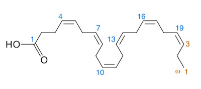
Docosahexaenoic acid (DHA), a component of fish oil, is a powerful therapeutic agent that can protect brain tissue and promote recovery in an experimental model of acute ischemic stroke, even when treatment is delayed by up to five hours.

These findings not only target a new stroke treatment approach, but also provide vital information about the length of the therapeutic window. The NIH-funded research is published in the journal, Translational Stroke Research, now available online at http://dx.doi.org/10.1007/s12975-010-0046-0.

Ischemic strokes result from loss of blood flow to an area of the brain due to a blockage such as a clot or atherosclerosis. The damage includes an irreversibly injured core of tissue at the site of the blockage. The area of tissue surrounding the core, called the penumbra, is also damaged but potentially salvageable.

The penumbra has a limited life span and appears to undergo irreversible damage within a few hours unless blood flow is reestablished and neuroprotective therapy is administered. A cascade of chemicals floods the tissue along with restored blood flow, including damaging free radicals and pro-inflammatory enzymes which can cause further damage and cell death.


DHA is an essential omega-3-fatty acid and is vital for proper brain function. It is also necessary for the development of the nervous system, including vision.

Moreover, omega-3 fatty acids, found in cold water fatty fish, including salmon, tuna, mackerel, sardines, shellfish, and herring, are part of a healthy diet that helps lower the risk of heart disease. DHA has potent anti-inflammatory effects.

Since inflammation is at the root of many chronic diseases, DHA treatment has been widely demonstrated to have beneficial effects in patients with coronary heart disease, asthma, rheumatoid arthritis, osteoporosis, sepsis, cancer, dry eye disease, and age-related macular degeneration, but its potential benefit in stroke was not known.

“We are just now beginning to understand the significant impact of omega-3 essential fatty acids on stroke,” notes Dr. Bazan. “There is no simple solution just yet, but each new discovery brings us closer to defeating stroke and other debilitating neurodegenerative diseases.”